Webmaster: LISI Dr Daniele Via Gaetano Mazzoni 29A - 00166 Roma ITALY - P.Iva 07049921005 mail postmaster@gastroatlas.org
I am aware that you will use these images for your presentations and I accept it, I would do the same.
At least thank the author of the report and the website
(example: courtesy of AUTHOR(S), see more on www.gastroatlas.org)
Sort by technique (Endoscopy, Ultrasound, etc.) or anathomical site
Free search text (e.g. by author, pathology, anathomical site, etc.)













1 2 3 4 › 4
Portal vein aneurysm EUS 737525
3 Ultrasound 5 EUS T MIscellaneous and other diseases 6 Radiology portal vein
Authors: LISI Daniele, LUCIDI Cristina, BIANCANIELLO Francesca, POMPA Valentina, VENTURINI Luigi, D’OVIDIO Valeria, Roma, Italy
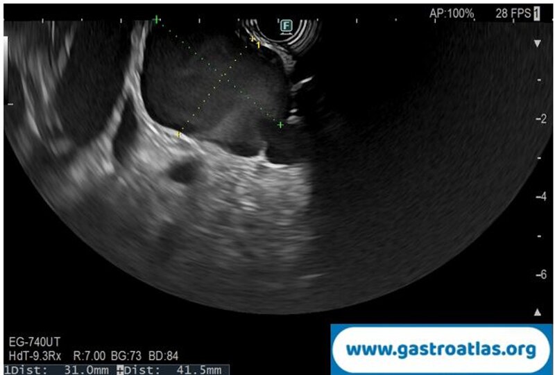
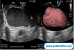
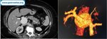
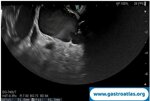
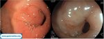
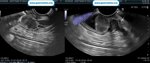
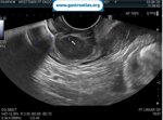
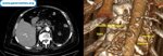
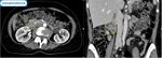
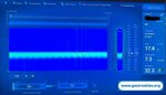
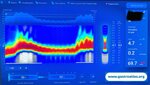
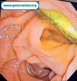
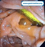
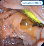
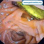
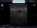
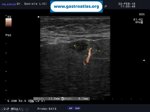
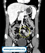
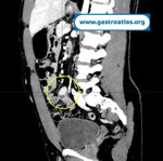
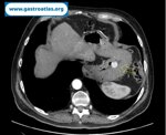
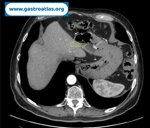
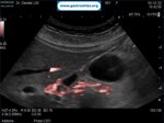
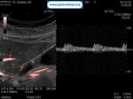
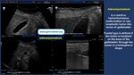
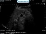
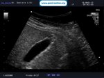
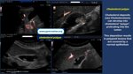
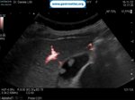
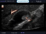
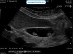
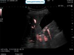
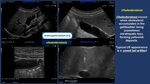
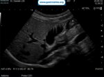
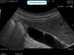
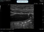
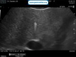
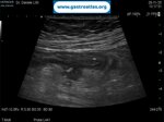
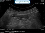
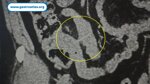
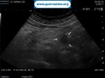
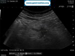
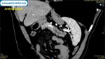
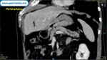
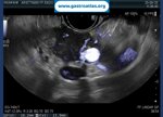
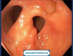
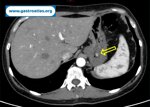
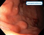
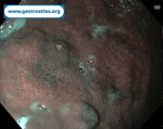
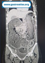
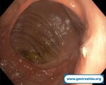
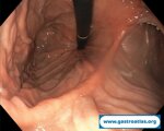
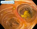
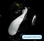
A 69-year-old woman was referred to our Unit for the evaluation of an incidental anechoic lesion near the hepatic hilum. A previous contrast-enhanced CT scan, performed two years earlier, had identified a vascular lesion compressing the pancreatic head. However, she was not referred for further investigation at that time. Endoscopic ultrasound (EUS) demonstrated a 42 × 31 mm anechoic lesion communicating with the portal vein, with regular walls and turbulent flow on e-flow and dual-DFI evaluation. Contrast-enhanced EUS confirmed turbulent flow synchronous with the portal venous phase, suggesting a portal vein aneurysm, subsequently confirmed by CT. Following multidisciplinary discussion, conservative management with clinical and radiological follow-up was recommended, considering the lesion's stability and the absence of symptoms over two years.
EUS video available https://www.e-ce.org/journal/view.php?number=8237